The following was originally published in Johns Hopkins Medicine’s Newsroom.
Johns Hopkins biomedical engineers have developed an artificial intelligence (AI) training strategy to capture images of mouse brain cells in action. The researchers say the AI system, in concert with specialized ultra-small microscopes, make it possible to find precisely where and when cells are activated during movement, learning and memory. The data gathered with this technology could someday allow scientists to understand how the brain functions and is affected by disease.
The researcher’s experiments in mice were published in Nature Communications on March 22.
“When a mouse’s head is restrained for imaging, its brain activity may not truly represent its neurological function,” says Xingde Li, Ph.D., professor of biomedical engineering at the Johns Hopkins University School of Medicine. “To map brain circuits that control daily functions in mammals, we need to see precisely what is happening among individual brain cells and their connections, while the animal is freely moving around, eating and socializing.”
Many of Li’s technologies are available for licensing through Johns Hopkins Technology Ventures.
To gather this extremely detailed data, Li’s team developed ultra-small microscopes that the mice can wear on the top of their head. Measuring in a couple of millimeter in diameter, the size of these microscopes limit the imaging technology they can carry on board. In comparison to benchtop models, the frame rate on the miniature microscopes is low, which make them susceptible to interference from motion. Disturbances such as the mouse’s breathing or heart rate would affect the accuracy of the data these microscopes can capture. Researchers estimate that Li’s miniature microscope would need to exceed 20 frames per second to eliminate all the disturbances from the motion of a freely moving mouse.
“There are two ways to increase frame rate,” says Li. “You can increase the scanning speed and you can decrease the number of points scanned.”
In previous research, Li’s engineering team quickly found they hit the physical limits of the scanner, reaching six frames per second, which maintained excellent image quality but was far below the required rate. So, the team moved on to the second strategy for increasing frame rate — decreasing the number of points scanned. However, similar to reducing the number of pixels in an image, this strategy would cause the microscope to capture lower-resolution data.
Li hypothesized that an AI program could be trained to recognize and restore the missing points, enhancing the images to a higher resolution. Such AI training protocols are used when it is impossible or time consuming to create a computer program for a task, such as reliably recognizing a cluster of features as a human face. Instead, computer scientists use the approach of letting computers learn to program themselves through processing large sets of data.
One significant challenge in the proposed AI approach was the lack of similar images of mouse brains to train the AI against. To overcome this gap, the team developed a two-stage training strategy. The researchers began training the AI to identify the building blocks of the brain from images of fixed samples of mouse brain tissue. They next trained the AI to recognize these building blocks in a head-restrained living mouse under their ultra-small microscope. This step trained the AI to recognize brain cells with natural structural variation and a small bit of motion caused by the movement of the mouse’s breathing and heartbeat.
“The hope was that whenever we collect data from a moving mouse, it will still be similar enough for the AI network to recognize,” says Li.
Then, the researchers tested the AI program to see if it could accurately enhance mouse brain images by incrementally increasing the frame rate. Using a reference image, the researchers reduced the microscope scanning points by factors of 2, 4, 8, 16 and 32 and observed how accurately the AI could enhance the image and restore the image resolution.
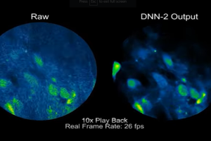
The researchers found that the AI could adequately restore the image quality up to 26 frames per second.
The team then tested how well the AI tool performed in combination with a mini microscope attached to the head of a moving mouse. With the combination AI and microscope, the researchers were able to precisely see activity spikes of individual brain cells activated by the mouse walking, rotating and generally exploring its environment.
“We could never have seen this information at such high resolution and frame rate before,” says Li. “This development could make it possible to gather more information on how the brain is dynamically connected to action on a cellular level.”
The researchers say that with more training, the AI program may be able to accurately interpret images up to 52 or even 104 frames per second.
Other researchers involved in this study include Honghua Guan, Dawei Li, Hyeon-cheol Park, Ang Li, Yungtian Gau and Dwight Bergles of the Johns Hopkins University School of Medicine; Yuanlei Yue and Hui Lu of George Washington University; and Ming-Jun Li from Corning Inc.
This research was supported by the National Cancer Institute (R01 CA153023), the National Science Foundation Major Research Instrumentation grant (CEBT1430030) and the Johns Hopkins Medicine Discovery Fund Synergy Award.